|
True Hermaphroditism [Images in Clinical Medicine] Karam, Jose A.; Baker, Linda A. University of Texas Southwestern Medical Center at Dallas; Dallas, TX 75390 |
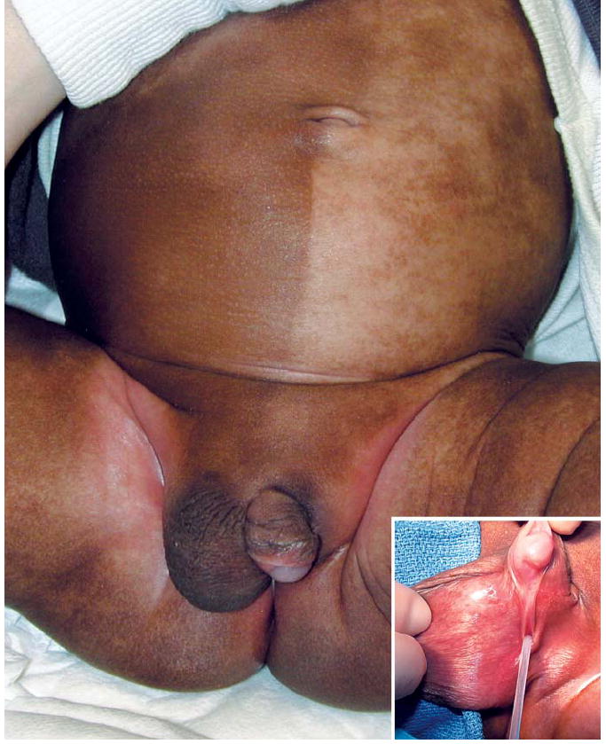
| A 17-day-old black neonate presented with ambiguous genitalia. A sharp line separated hypopigmented skin on the left side of the abdomen from normally pigmented skin on the right side. A pendulous, wrinkled labioscrotal fold contained a gonad on the right side. In contrast, the hypoplastic, flat, left side was empty. A palpable left inguinal mass could not be clearly characterized with the use of ultrasonography. The phallus was 3 cm in length, with perineal hypospadias (inset) and 30-degree chordee. Testing revealed 46,XX/46,XY mosaicism and normal levels of male hormones. Laparoscopic surgery was performed; on the left side a hemiuterus, a fallopian tube, fimbriae, and an ovary were found and resected. These structures were absent on the right side. A normal vas exited the right internal ring, leading to a well-formed epididymis and a scrotal, biopsy-proven ovotestis with dotted islands of ovarian tissue capping the lower pole of the testicle. The ovarian tissue was removed, and left inguinal herniorrhaphy, placement of a left testicular prosthesis, and repair of the hypospadias were subsequently completed. At 21 months, the child has an excellent cosmetic result. |